This image has twenty-seven channels and your figure has three
Imaging mass cytometry records one channel per antibody. A page holds three colours before they stop being tellable apart, and even the three that cover the most leave half of what was stained unshown.
A fluorescence microscope runs out of colours at about four. The dyes overlap, the filters bleed, and past that nothing can be told from anything else. So a panel is four markers, and a question needing a fifth needs a second experiment.
Imaging mass cytometry does not work that way. Each antibody carries a different metal isotope, and the instrument counts the metals by mass, so the channels cannot bleed into one another at all. This section of human tonsil was recorded with 27.
The page is still flat and still has three colours.

-
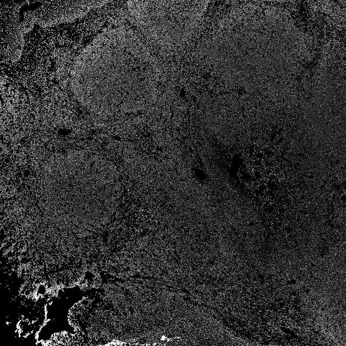
The nuclear counterstain. Every cell in the section, which is what makes it the right thing to measure the others against: a marker covering a tenth of the section and a marker covering a tenth of the cells are different claims.
DNA1-191/193Ir, marking 86.6% of the tissue.
-
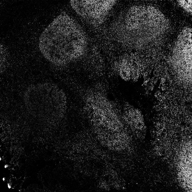
CD19, a B cell marker. The follicles appear, because that is what a follicle is made of.
CD19-169Tm, marking 8.6% of the tissue.
-
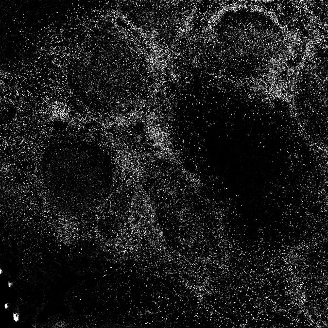
CD3, a T cell marker. It lights the spaces between the follicles, almost exactly where the last panel was dark. Two channels, two populations, one section.
CD3-170Er, marking 16.1% of the tissue.
-
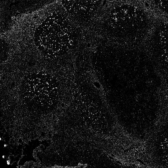
CD68, found on macrophages. Not a region this time but a scatter of individual cells, present in both compartments and belonging to neither.
CD68-171Yb, marking 5.4% of the tissue.
-
And the blank. No antibody, carried through the run anyway. This is what the instrument records when there is nothing there, and it is the only reason to believe the panels above it.
empty, marking 1.9% of the tissue.
Five of the twenty-seven, one at a time
Each panel is one antibody, shown on its own. The last is the panel's own blank: an empty channel carried through the run, which is what the instrument records where there is nothing to find. It marks 1.86% of the tissue, and it is the thing to check any claim about the other channels against.
Those four markers divide the section in ways that have nothing to do with one another. B cell regions, T cell regions, a population scattered through both. A figure showing any one of them is true and is not the tissue.
What three colours hold
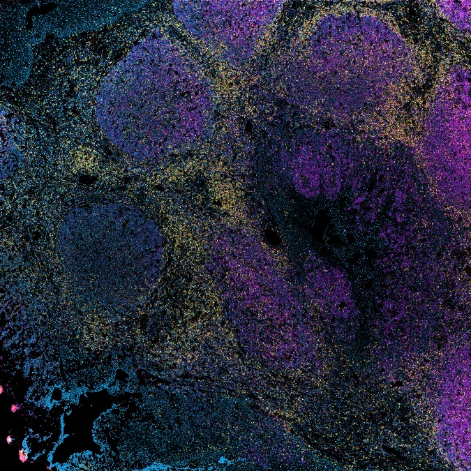
Three channels of the twenty-seven
DNA, CD3 and CD19 laid over one another. We chose these three and the colours are this site's, not the archive's: it is the view this tissue is usually shown in, and it is a good figure. It is also three of the 27 channels that were recorded.
That is a good figure and this piece is not an argument against it. The question is what it costs, and that can be counted.
Setting aside the two nuclear channels, which mark every cell by design, and the blank, there are 24 markers. Between them they mark 85.1% of the tissue in the section.
Take the three markers that between them cover the most, found greedily rather than chosen. They are CD185, CD1c, CD370, and they cover 50.2% of everything the 24 markers mark.
That is a ceiling and not an estimate. Any three picked for being informative rather than for being large cover less than that, including the three in the figure above. Half, at best, of what the instrument was asked to find.
It takes 11 channels to reach 90%. Not three, not four, and not a number a page can carry.
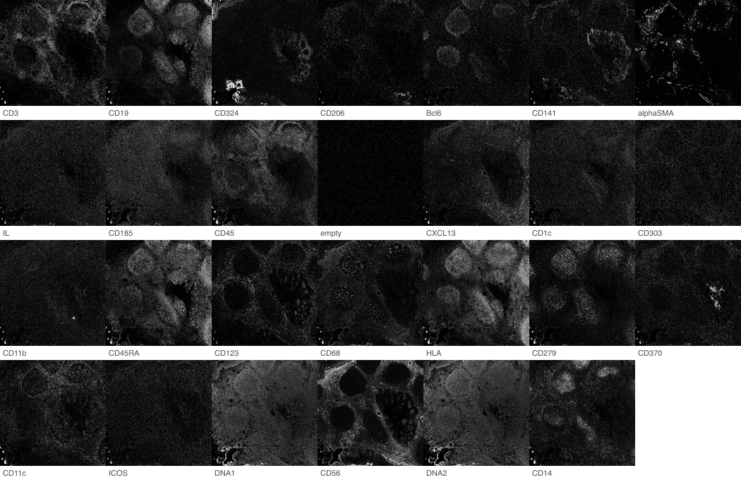
Every channel in the file, in the order it is stored, each on its own and each scaled to its own range. The blank is the dark one in the second row. Measured at pyramid level 2 of 5, which is 675 by 675 of the 2700 by 2702 recorded.
| Channel | Share of tissue marked |
|---|---|
| DNA2-191/193Ir (counterstain) | 90.61% |
| DNA1-191/193Ir (counterstain) | 86.63% |
| CD185/CXCR5-151Eu | 18.99% |
| CD1c/BDCA1-biotin + Neutravidin-173Yb | 17.60% |
| CD45RA-155Gd | 17.46% |
| CD370/Clec9A-161Dy | 16.22% |
| CD3-170Er | 16.08% |
| CD11b-149Sm | 15.63% |
| HLA-DR-174Yb | 15.03% |
| Bcl6-163Dy | 14.36% |
| CD14-156Gd | 12.93% |
| CD45-152Sm | 12.71% |
| IL-21-164Dy | 11.81% |
| ICOS-148Nd | 11.03% |
| CD123-143Nd | 10.55% |
| CD19-169Tm | 8.58% |
| CD141/BDCA3-165Ho | 7.67% |
| CXCL13-157Gd | 7.47% |
| CD279/PD-1-175Lu | 7.28% |
| CD324/E-Cadherin-158Gd | 6.23% |
| CD303/BDCA2-147Sm | 6.06% |
| CD56-176Yb | 6.01% |
| CD206-168Er | 5.81% |
| CD68-171Yb | 5.36% |
| CD11c-159Tb | 4.74% |
| empty (blank) | 1.86% |
| alphaSMA-141Pr | 1.12% |
The blank is the reason to believe any of this
Every number here rests on deciding, per channel, which pixels hold signal. That is a threshold, and a threshold can be moved until the answer is the one you wanted.
The panel carries its own control. One channel had no antibody in it and was carried through the run regardless, and whatever method is used to say how much a channel found has to say that this one found almost nothing. It ranks 26 of 27 and marks 1.86% of the tissue. The script that produced these figures refuses to write them if that stops being true.
That is not a flourish. It is the difference between a measurement and a number, and it was in the authors’ data rather than ours: they put the blank in the panel because they intended somebody to check.
What to do with twenty-seven channels and three colours
Nothing here says publish a twenty-seven panel figure. Nobody would read it, and the grid above is legible only because it is making an argument about itself rather than about tonsils.
What is worth saying is smaller. The figure is a selection, the selection is an argument, and the reader cannot see which twenty-four things were set aside. Saying how many channels were recorded costs one clause in a legend. Depositing the rest, which these authors did, costs nothing at all and is why this page could be written by somebody who has never touched the tissue.